Abdominal Anatomy - Anatomy Of The Abdomen And Pelvis A Journey From Basis To Clinic Coursera. These two apertures, together with abdominal walls, bound the abdominal cavity. Together, these three turn nutrients into usable energy, as well as help dispose of solid waste. Observation, auscultation, percussion, and palpation. Terms in this set (94) what is the abdomen. Posterior abdominal wall edit posterior wall muscles edit source.
These organs are held together loosely by connecting tissues. Part of the trunk between thorax and pelvis. We're going to take apart a plastic anatomy model and see what we can find in the abdomen. To reach this goal and minimize complications, every reproductive surgeon requires a thorough knowledge of pelvic anatomy. Abdominal muscle strains don't cause a bulge or visible lump.
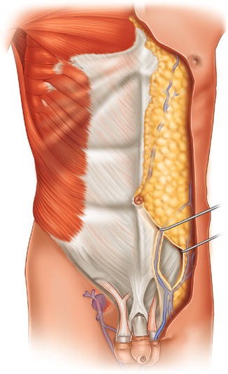
Anatomical Illustration Of The Abdominal Wall And Fat Springerlink from media.springernature.com Its superior aperture faces towards the thorax, enclosed by the diaphragm. These organs are held together loosely by connecting tissues. For the sake of brevity, the various organs will be not discussed in detail. Abdominal wall anatomy that is clinically pertinent to the surgeon, focusing primarily on the structures of the anterior abdominal wall, will be reviewed. The region occupied by the abdomen is called the abdominal cavity, and is enclosed by the abdominal muscles at front and to the sides, and by part of the vertebral column at the back. Quadratus, quadrate shape on the lateral side of the posterior abdominal wall. The goal, whenever possible, is to restore normal anatomy. We're going to take apart a plastic anatomy model and see what we can find in the abdomen.
Related posts of anatomy of the abdomen women anatomical parts of the digestive system.
The rectus abdominis connects to the xiphoid process, a bony landmark at the bottom of the sternum. The abdominal wall surrounds the abdominal cavity, providing it with flexible coverage and protecting the internal organs from damage. Inferiorly the abdomen is open to the pelvis, communicating through the superior pelvic aperture (pelvic inlet). Abdominal muscle strains don't cause a bulge or visible lump. The normal anatomy or organs imaged in a standard abdominal examination is explained below. The abdominal portion of the aorta supplies most of the abdomen, and begins at the level of the twelfth thoracic vertebra (t12), and then terminates at l4 by bifurcating into the left and right common iliac arteries. These two apertures, together with abdominal walls, bound the abdominal cavity. The aorta is the largest blood vessel in the body. Anatomical parts of the digestive system 12 photos of the anatomical parts of the digestive system anatomical description of digestive system, anatomical structure of the digestive system, anatomy and function of the digestive system, anatomy function of the digestive system, what are the anatomical. One of the easiest ways to tell if your pain is caused by a hernia or pulled stomach muscle is if you have a bulge or not. Common incisions and closure techniques, and prevention and management of wound complications, are discussed elsewhere. These organs are held together loosely by connecting tissues. The abdominal aorta enters the abdomen through the diaphragm at the level of the twelfth thoracic vertebre and continues to just below the umbilical area, where it splits into the right and left common iliac arteries.
Posterior abdominal wall edit source posterior wall muscles edit . The region occupied by the abdomen is called the abdominal cavity, and is enclosed by the abdominal muscles at front and to the sides, and by part of the vertebral column at the back. The rectus abdominis connects to the xiphoid process, a bony landmark at the bottom of the sternum. These organs are held together loosely by connecting tissues. The abdomen (colloquially called the belly, tummy, midriff or stomach) is the part of the body between the thorax (chest) and pelvis, in humans and in other vertebrates.
Female Abdominal Anatomy Images Koibana Info Human Body Organs Human Body Anatomy Body Anatomy from i.pinimg.com Auscultation before percussion) and carry different degrees of importance. Boundaries of the abdomen (4) anterior abdominal wall (anterolateral) diaphragm (superior) The abdominal portion of the aorta supplies most of the abdomen, and begins at the level of the twelfth thoracic vertebra (t12), and then terminates at l4 by bifurcating into the left and right common iliac arteries. The region occupied by the abdomen is called the abdominal cavity, and is enclosed by the abdominal muscles at front and to the sides, and by part of the vertebral column at the back. The normal anatomy or organs imaged in a standard abdominal examination is explained below. Abdomen anatomy the abdomen is comprised primarily of the digestive tract and other accessory organs which assist in digestion, the urinary system, spleen, and the abdominal muscles (shown below). Abdominal wall anatomy that is clinically pertinent to the surgeon, focusing primarily on the structures of the anterior abdominal wall, will be reviewed. Terms in this set (94) what is the abdomen.
Inferiorly the abdomen is open to the pelvis, communicating through the superior pelvic aperture (pelvic inlet).
Common incisions and closure techniques, and prevention and management of wound complications, are discussed elsewhere. The area occupied by the abdomen is called the abdominal cavity. Abdominal computed tomography (ct) is a type of medical imaging procedure used to diagnose and monitor internal stomach issues, like cancer, bowel obstruction, and abdominal pain. One of the easiest ways to tell if your pain is caused by a hernia or pulled stomach muscle is if you have a bulge or not. Stomach is a muscular bag forming the most distensible part of the human digestive system. The region occupied by the abdomen is called the abdominal cavity, and is enclosed by the abdominal muscles at front and to the sides, and by part of the vertebral column at the back. To reach this goal and minimize complications, every reproductive surgeon requires a thorough knowledge of pelvic anatomy. A hernia will usually cause a distinct bulge where the tissue or organ pushes through the muscle wall. The abdomen contains all the digestive organs, including the stomach, small and large intestines, pancreas, liver, and gallbladder. Quadratus, quadrate shape on the lateral side of the posterior abdominal wall. Its superior aperture faces towards the thorax, enclosed by the diaphragm. For the sake of brevity, the various organs will be not discussed in detail. The majority of these organs are encased in a protective membrane termed the peritoneum.
It is the long, flat muscle that extends vertically between the pubis and the fifth, sixth, and seventh ribs. The major components of the abdominal exam include: The aorta is the largest blood vessel in the body. Abdomen anatomy the abdomen is comprised primarily of the digestive tract and other accessory organs which assist in digestion, the urinary system, spleen, and the abdominal muscles (shown below). The abdomen is the body region found between the thorax and the pelvis.
Internal Anatomy Of Male Chest And Abdomen On Black Stock Photo Download Image Now Istock from media.istockphoto.com Abdomen anatomy the abdomen is comprised primarily of the digestive tract and other accessory organs which assist in digestion, the urinary system, spleen, and the abdominal muscles (shown below). We'll identify as many organs as we can, see how they fit into the. The abdomen is the front part of the abdominal segment of the trunk. The coeliac trunk arises from the abdominal aorta at t12 and supplies the foregut gastrointestinal viscera. These organs are held together loosely by connecting tissues. Inferiorly the abdomen is open to the pelvis, communicating through the superior pelvic aperture (pelvic inlet). For the sake of brevity, the various organs will be not discussed in detail. The normal anatomy or organs imaged in a standard abdominal examination is explained below.
These two apertures, together with abdominal walls, bound the abdominal cavity.
Together, these three turn nutrients into usable energy, as well as help dispose of solid waste. The aorta is the largest blood vessel in the body. Abdominal muscle strains don't cause a bulge or visible lump. We're going to take apart a plastic anatomy model and see what we can find in the abdomen. The normal anatomy or organs imaged in a standard abdominal examination is explained below. The abdomen is the part of the body that contains all of the structures between the thorax (chest) and the pelvis, and is separated from the thorax via the diaphragm. The abdomen contains all the digestive organs, including the stomach, small and large intestines, pancreas, liver, and gallbladder. The diaphragm marks the top of the abdomen and the horizontal line at the level of the top of the pelvis marks the bottom. While these are the same elements which make up the pulmonary and cardiac exams, they are performed here in a slightly different order (i.e. The abdomen is the front part of the abdominal segment of the trunk. Related posts of anatomy of the abdomen women anatomical parts of the digestive system. It is an artery, meaning that it carries blood away from the heart. Posterior abdominal wall edit posterior wall muscles edit source.
Share :
Post a Comment
for "Abdominal Anatomy - Anatomy Of The Abdomen And Pelvis A Journey From Basis To Clinic Coursera"


Post a Comment for "Abdominal Anatomy - Anatomy Of The Abdomen And Pelvis A Journey From Basis To Clinic Coursera"